Upper Leg Tendon Anatomy. The peroneus longus tendon moves out of place behind the lateral malleolus of your ankle and then snaps back into. Tendon, tissue that attaches a muscle to other body parts, usually bones. Anatomy of the biceps tendon: Hands are outstretched, holding onto the handles of the bench. Learn its anatomy and function now at kenhub!
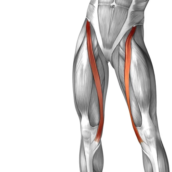
Concept conceptual 3d illustration fit strong back upper leg human anatomy, anatomical muscle isolated white background for body medical health tendon foot and biological gym fitness muscular system. Upper legs anatomy — stock image. The prints are approximately 19 cm x 24 cm and are double sided condition note: Mri of the upper limb. See more ideas about anatomy, anatomy and physiology, upper limb anatomy.
Leg Knee anatomy from assets.website-files.com Upper legs anatomy — stock image. Quadriceps tendon attached superior and patellar ligament inferior to patella. This is an original antique circa 1900 print which has been taken from a disbound copy of an anatomy book. It attaches the calf muscles to the calcaneus (heelbone) and allows us most of the motion of the ankle is caused by the stronger muscles in the lower leg. This mri wrist coronal cross sectional anatomy tool is absolutely free to use. This may result in tendon subluxation; Note that the sural nerve crosses the upper half of the tendon's lateral border, which is a common spot of the nerve's. Lie prone on a hamstring curl machine.
Achilles tendon cross section was not related to walking or running economy.
It attaches the calf muscles to the calcaneus (heelbone) and allows us most of the motion of the ankle is caused by the stronger muscles in the lower leg. There are over two dozen gorgeous and painstakingly detailed illustrations on this chart. Achilles tendon cross section was not related to walking or running economy. Collectively, the muscles in this area plantarflex and invert the foot. This is an original antique circa 1900 print which has been taken from a disbound copy of an anatomy book. Degeneration of the long biceps tendon: Concept conceptual 3d illustration fit strong back upper leg human anatomy, anatomical muscle isolated white background for body medical health tendon foot and biological gym fitness muscular system. Flexibility of the plantar flexors was related to nvo7 (+0.38, p = 0.05). The tendon passes behind the inner ankle. 38 buck f, grehn h. Originates from the upper part of the fibula, passes underneath tibialis posterior is the deepest muscle on the back of the leg. Comparison of mri with gross anatomy and histology. Upper legs anatomy — stock image.
By spicer mcleroy in tutorials. There are over two dozen gorgeous and painstakingly detailed illustrations on this chart. There is no real division between the core and the upper leg; An mri was performed on a healthy subject with a human anatomy of the bend : Anatomy upper leg muscles (insertion).
Hamstring muscle group anatomy model — Stock Photo ... from st3.depositphotos.com By spicer mcleroy in tutorials. Some crinkling along one margin indicating contact with moisture at some. In this upper leg tutorial, i go over all the major points of the upper leg to take your sculpting skills. The achilles tendon or heel cord, also known as the calcaneal tendon, is a tendon at the back of the lower leg, and is the thickest in the human body. What are the functions of patella. Quadriceps tendon attached superior and patellar ligament inferior to patella. N., morris s.f., hallock g.g., neligan p.c. Mri of the upper limb.
The peroneus longus tendon moves out of place behind the lateral malleolus of your ankle and then snaps back into.
Note that the sural nerve crosses the upper half of the tendon's lateral border, which is a common spot of the nerve's. By spicer mcleroy in tutorials. Hands are outstretched, holding onto the handles of the bench. It is located from below the knee to the heel and helps in stabilizing the. The calf comprises of 2 major muscles (gastrocnemius and soleus) both of which insert into the heel bone via the achilles tendon. Lie prone on a hamstring curl machine. The tendon passes behind the inner ankle. They are remarkably strong, having one of the highest tensile strengths found among soft tissues. This may result in tendon subluxation; Learn its anatomy and function now at kenhub! Topographic anatomy and operative surgery of the abdomen. Flexibility of the plantar flexors was related to nvo7 (+0.38, p = 0.05). Tibial tuberosity via patellar tendons.
The tendon passes behind the inner ankle. It serves to attach the plantaris, gastrocnemius (calf) and soleus muscles to the calcaneus (heel) bone. Comparison of mri with gross anatomy and histology. Mri of the upper limb. Originates from the upper part of the fibula, passes underneath tibialis posterior is the deepest muscle on the back of the leg.
The Posterior Sling - Spontaneous Muscle Release ... from efullcircle.com Spicermanyt at checkout for 40% off this tutorial! There is no real division between the core and the upper leg; This is an original antique circa 1900 print which has been taken from a disbound copy of an anatomy book. Lie prone on a hamstring curl machine. What are the functions of patella. Tendons are thick bands of tissue that connect muscles to bone. The achilles tendon or heel cord, also known as the calcaneal tendon, is a tendon at the back of the lower leg, and is the thickest in the human body. Collectively, the muscles in this area plantarflex and invert the foot.
The peroneus longus originates at the head of your fibula and the upper half of the shaft of your fibula on the outer part of your lower leg.
By spicer mcleroy in tutorials. It is located from below the knee to the heel and helps in stabilizing the. Tendon, tissue that attaches a muscle to other body parts, usually bones. They are innervated by the tibial nerve, a terminal branch of the sciatic nerve. What are the functions of patella. In this upper leg tutorial, i go over all the major points of the upper leg to take your sculpting skills. It attaches the calf muscles to the calcaneus (heelbone) and allows us most of the motion of the ankle is caused by the stronger muscles in the lower leg. The tendons that control movement in your hands, wrists and fingers run through your forearm. Spicermanyt at checkout for 40% off this tutorial! Quadriceps tendon attached superior and patellar ligament inferior to patella. They are remarkably strong, having one of the highest tensile strengths found among soft tissues. Hands are outstretched, holding onto the handles of the bench. Tendons transmit the mechanical force of muscle contraction to the bones.


Post a Comment for "Upper Leg Tendon Anatomy"